Brain and Arteries
How a stroke affects you depends on where in the brain it occurs. It’s possible for a stroke to affect how you think, as well as the way you move, how your senses work, or how you use language.
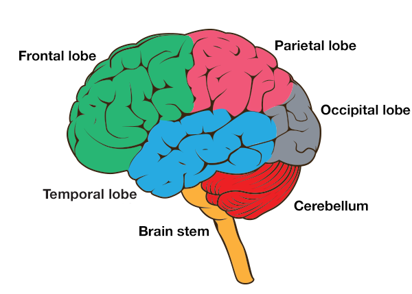
- Frontal lobe: Located behind the forehead, this is the front section of the cerebrum, which is the large, wrinkled part of the brain that looks like a walnut and is where most thinking occurs. Strokes in the frontal lobe can affect a person’s ability to plan, concentrate and solve problems. They can also lead to changes in personality, initiative and emotions, or affect the control of muscles in the legs, arms and hands, and muscles used for speaking and swallowing.
- Parietal lobe: The part of the cerebrum at the top rear of the skull, it is important for being aware of your surroundings. It does this through spatial awareness, the senses of touch, pain and feeling hot or cold, feeling where your body and limbs are without needing to look (proprioception), and calculation and writing.
- Temporal lobe: Part of the cerebrum at the side of the head, near your temples and ears. It plays a big role in understanding speech and other sounds, and your recognition of faces and objects.
- Occipital lobe: The part of the cerebrum at the back of the skull, it is devoted to vision.
- Cerebellum: Situated at the rear of the brain below the cerebrum, it controls your balance and the coordination of muscles. Strokes in this area can cause nausea and headaches, and affect your posture and how you walk.
- Brain stem: The part of the brain that connects to the spinal cord, it controls automatic functions like heart rate, breathing, blood pressure, sweating, and movement of your pupils, eyes and face. Strokes here can affect many parts of the body and senses, and cause vertigo, dizziness and imbalance. In rare cases a brain stem stroke can lead to locked-in syndrome.
Deep inside the brain are other structures that can’t be seen on the above diagram. These include the midbrain, which controls whether you’re asleep or awake; the thalamus, which combines sensory information to form a picture of the world; the hypothalamus, which controls hormones and internal organs; the hippocampus, which forms new long-term memories; the amygdala, which controls emotional responses; and the basal ganglia, which are involved in fine body movements, learning and habit formation.
Nerves from the left and right side of the body cross over in the brain stem. Because of this, the location of the stroke in the brain usually affects the opposite side of the body.
The left hemisphere or half of the brain controls most functions on the right side of the body, while the right hemisphere controls most functions on the left side.
Blood supply to the brain
The two types of stroke both involve an interruption to the brain’s blood supply, which is carried in blood vessels called arteries. An interruption may happen when an artery is blocked, or when it bursts and bleeds.
The brain cells die when they don’t get enough blood and miss out on the oxygen and important nutrients that the blood carries. Some of these cells die shortly after the stroke starts, but some can last a few hours if the blood supply is not cut off completely. They might be able to recover if blood supply is returned in the minutes and hours after the stroke.
The part of the brain damaged by the stroke is determined by which artery is affected.
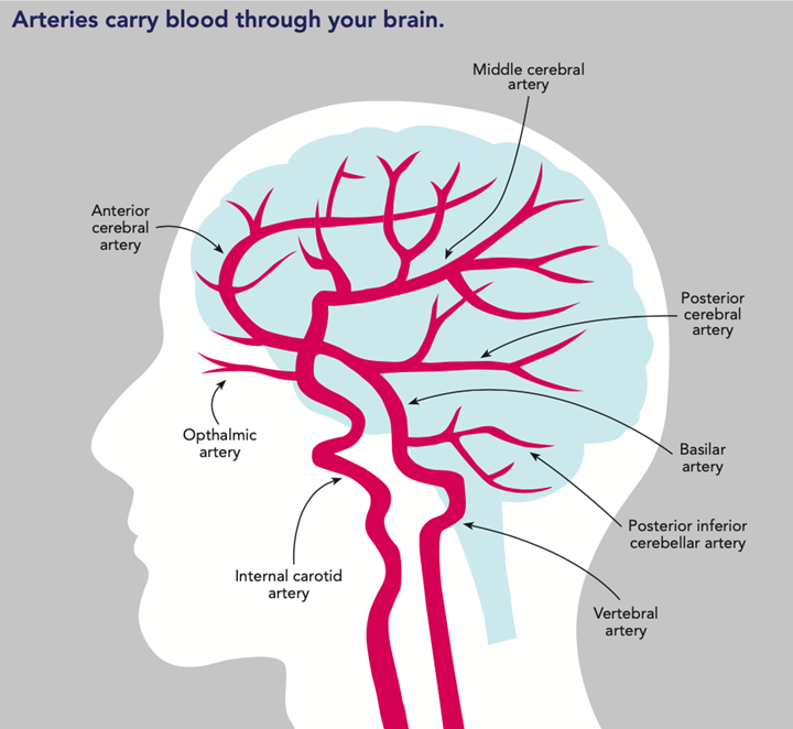
- Anterior cerebral artery: Supplies blood to the areas of the frontal lobe and the top of the parietal lobe that are close to the divide between the two hemispheres. There are actually two anterior cerebral arteries, one each for the left and right hemispheres.
- Middle cerebral artery: One of a pair of arteries that branch out to supply blood to the left and right sides of the brain, mostly to the temporal lobes and parts of the frontal and parietal lobes.
- Posterior cerebral artery: One of a pair of arteries that supply blood to the occipital lobe, the bottom rear parts of the temporal lobes, and the thalamus.
- Ophthalmic artery: One of a pair of arteries that supply blood to the eyes and some of the muscles around the eyes and nose.
- Basilar artery: A single artery that begins where the two vertebral arteries join. Branches off the basilar artery supply blood to the cerebellum, parts of the brain stem and the inner ear. However, most of the blood goes up to the circle of Willis, which is where it joins with the internal carotid arteries to supply the anterior, middle and posterior cerebral arteries.
- Internal carotid artery: One of a pair of arteries, running on the left and right sides of the neck. They are the biggest arteries that go into the brain, and are where you can feel the pulse of the heartbeat lower down in the neck. In the brain they form the circle of Willis with the basilar artery, and supply blood to the anterior, middle and posterior cerebral arteries.
- Vertebral artery: One of two arteries that run up through the vertebrae or bones in the neck. The vertebral arteries join to form the basilar artery, and they also supply blood to part of the cerebellum.
A stroke affects your brain.
It changes how your brain works.
What it does depends on where it is in the brain.
Each part of the brain has a different job.
The frontal lobe is at the front of the brain.
You use it to think.
A stroke in the frontal lobe can affect:
The parietal lobe is at the top of the brain.
You use it to know what is around you.
You use it feel where parts of your body are without looking.
A stroke in the parietal lobe can affect:
The temporal lobe is at the side of the brain.
You use it to hear.
You use it to know a face when you see it.
A stroke in the temporal lobe can affect:
The occipital lobe is at the back of brain.
You use it to see.
A stroke in the occipital lobe can affect seeing.
The cerebellum is at the bottom of the brain.
You use it to balance.
You use it for coordination.
A stroke in the cerebellum can affect:
- walking
- headaches
- feeling sick.
The brain stem joins the brain to the body.
It is in charge of your heart and lungs.
A stroke in the brain stem can affect:
- the whole body
- senses
- balance.
There are other parts inside the brain:
- The midbrain is in charge of sleep.
- The thalamus combines senses.
- The hypothalamus is in charge of your organs.
- The hippocampus makes memories.
- The amygdala controls emotion.
- The basal ganglia helps you learn and move.
Sides of the brain
The brain has a left side and a right side.
The two sides are called hemispheres.
Each side of the brain controls the other side of the body.
The left side of the brain controls the right side of the body.
The right side of the brain controls the left side of the body.
Blood supply to the brain
Blood is carried to the brain in arteries.
A stroke happens when blood does not get to the brain.
This happens when an artery:
- is blocked
- bursts or bleeds.
Find out more about the types of stroke.
The brain is made out of brain cells.
Brain cells die if they do not get blood.
Some cells die straight away.
Some cells get better when blood gets back to the brain.
Each part of the brain gets blood from a different artery:
- Anterior cerebral artery takes blood to the front and top of the brain.
It goes near where the two hemispheres join. - Middle cerebral artery takes blood to the side of the brain.
- Posterior cerebral artery takes blood to the back of the brain.
- Ophthalmic artery takes blood to the eyes.
- Basilar artery takes blood to the cerebellum and brain stem.
It joins to the other arteries in the brain.
- Internal carotid artery takes blood up from the neck to the brain.
It joins to the other arteries in the brain. - Vertebral artery takes blood through the neck bones to the brain.
It joins to the basilar artery.